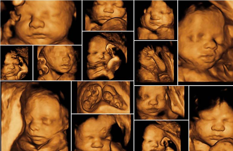
Gynec Sonography (3D and 4D)
Traditional sonography does not provide the same level of detail as 3-Dimensional or 4-Dimensional ultrasound. The 3D and 4D ultrasounds are optional and are not included in standard prenatal testing. If the parents are interested, the doctors will recommend 3D and 4D ultrasound scans. If the doctor suspects any birth defects, such as cleft palate or other structural birth anomalies, this type of ultrasonography is recommended.
3D and 4D ultrasounds, like traditional ultrasound procedures, use sound waves to create an image of the baby in the womb. The difference between 3D and 4D ultrasound is that 3D images are three-dimensional still images of the baby, whereas 4D images show real-time movements such as facial features, limbs, and body in detail, as well as the baby's expressions. The images from the 4D scan align in such a way that they appear to be a video.
Sound waves in 3D and 4D ultrasonography are directed at various angles, as opposed to 2D ultrasound, which is directed at a single angle. The sophisticated software generates images from sound waves reflected off of various surfaces. Surface rendering is a technique used in 3D and 4D ultrasound procedures. Except for the dimension of motion, 3D and 4D ultrasound procedures are similar.
The best time to get a 3D or 4D ultrasound scan is between 26 and 30 weeks of pregnancy. The baby has very little fat under the skin and the bones are clearly visible at 26 weeks of pregnancy. The visibility of the baby's face is also affected by the direction the baby is facing within the womb.
